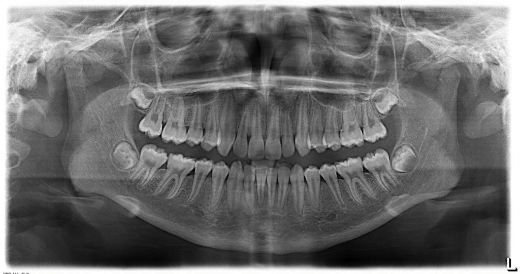
2D OPG
An orthopantamogram (OPG) or dental panoramic radiopgraph is a panoramic scanning dental radiograph (x-ray)
of the upper and lower jaw. OPG shows a two-dimensional view of both the jaws from ear to ear.Besides screening procedures, the most common use is to determine the status of wisdom teeth and trauma to the jaws.
With our technology, we offer high resolution panoramic radiographs.
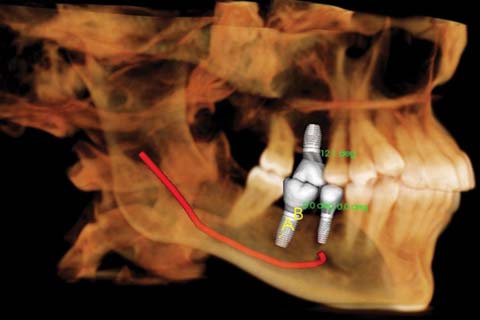
DENTAL IMPLANT PLANNING
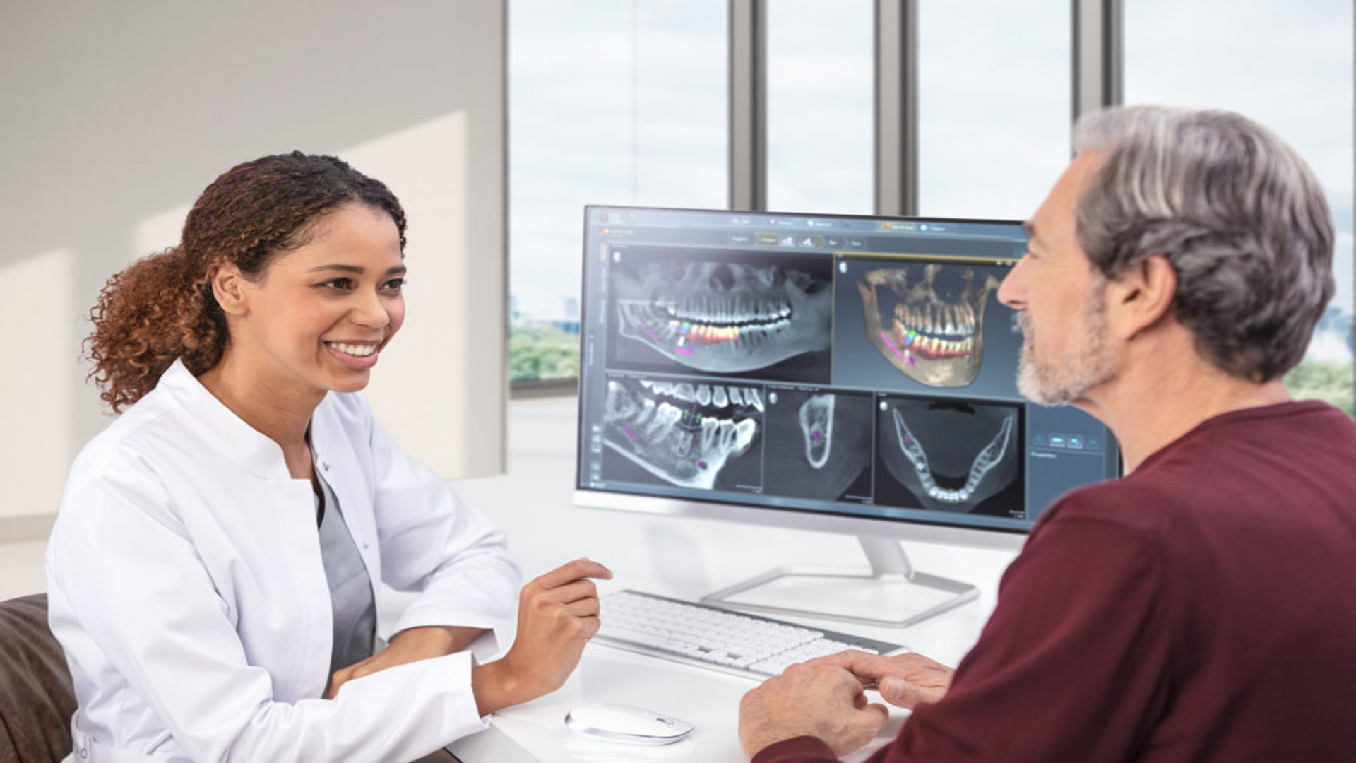
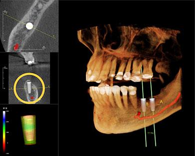
CBCT scans provide detailed 3D images of the jawbone, allowing for precise implant placement planning. This level of detail is not possible with traditional 2D imaging, such as x-rays.CBCT imaging can identify the location and shape of anatomical structures such as nerves, blood vessels, and sinuses, which is critical for identifying any potential complications or risks associated with implant placement.CBCT imaging can aid in treatment planning for dental implants, such as determining the optimal implant size, angulation, and placement to ensure long-term success.CBCT scans are an essential tool for accurate and safe dental implant planning, ensuring that the implant procedure is successful and minimizes potential complications.
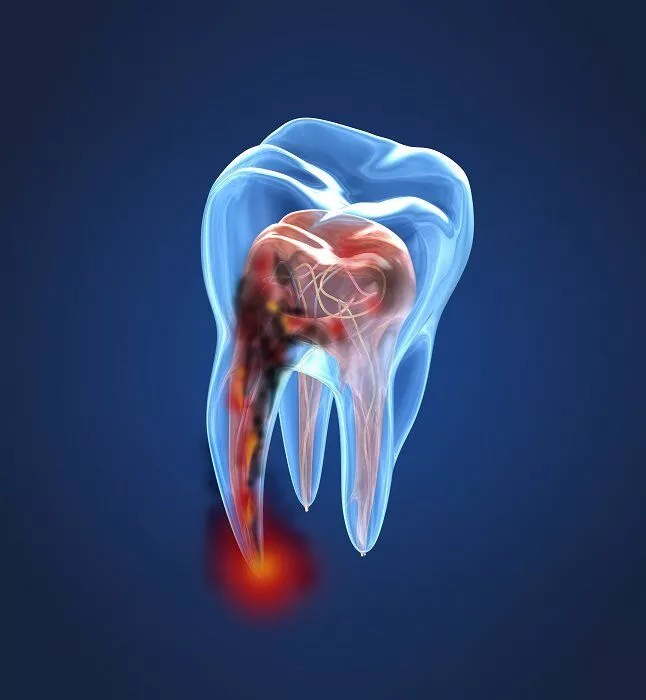
ENDODONTIC IMAGING
Cone Beam Computed Tomography (CBCT) is a valuable tool in endodontic imaging that provides detailed 3D images for identifying the anatomy of the root canal system, detecting root fractures, and assessing the extent of periapical lesions, the teeth and surrounding structures.CBCT imaging can aid in treatment planning for endodontic cases, such as determining the number and location of canals, identifying any calcified canals, and identifying any complications or risks associated with the root canal procedure.CBCT scans are an essential tool for accurate diagnosis and successful treatment of endodontic cases, ensuring that patients receive the best possible outcomes for their dental health.

ENT DR. REFERALS
CBCT scans provide detailed 3D images of the head and neck, allowing for precise diagnosis and treatment planning. This level of detail is not possible with traditional 2D imaging, such as x-rays.CBCT scans are an essential tool for accurate diagnosis, treatment planning, and surgical execution in ENT cases. By providing ENT doctors with a detailed view of the patient's anatomy, CBCT imaging can lead to improved patient outcomes and better quality of life.
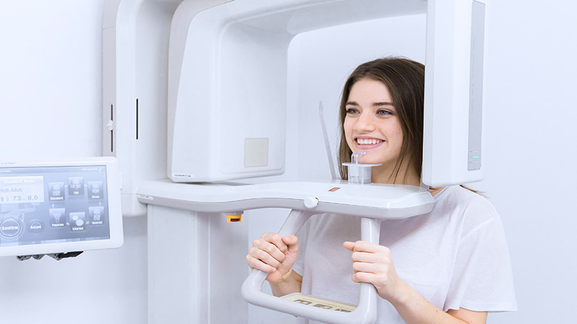
HI RES DENTAL CT SCANS
For many years dental professionals have relied on intraoral radiography, panoramic radiography, and conventional tomography for diagnosis and treatment planning. These commonly used imaging modalities produce only two-dimensional and/or distorted images with superimposition of structures outside the area of interest. There has always been a need for three-dimensional (3-D) imaging, but the technology has only recently become readily available. Cone Beam Computed Tomography (CBCT) uses a cone-shaped beam and digital processing to reconstruct a virtually distortion-free 3-D image using a single rotation in a sitting/standing position, similar to that of a panoramic radiograph. CBCT scanners are compact in size, capable of producing high-resolution 3-D imaging of hard tissues, and are compatible with computer-aided imaging software for improved diagnosis and treatment planning
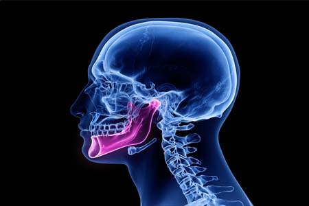
TMJ SCAN
Temporomandibular joint (TMJ) Cone Beam Computed Tomography (CBCT) scans are important for diagnosing and treating TMDs. CBCT imaging provides detailed 3D images of the TMJ, including the joint's anatomy, bone changes, and the position of the condyle. This level of detail is not possible with traditional x rays. CBCT imaging can aid in accurate diagnosis of TMJ disorders, which can be difficult to diagnose using clinical examination alone. This can lead to more effective treatment planning and better patient outcomes.